An animal eye diagram is a labeled illustration that shows the anatomical structures of eyes across different species, revealing how vision works through the physical arrangement of parts like the cornea, lens, retina, and optic nerve. These visual tools help us understand not just what makes up an eye, but how different creatures have evolved remarkably varied solutions to the challenge of seeing their world.
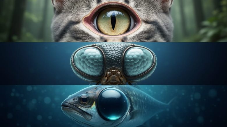
Whether you’re studying the compound eyes of an insect, the reflective tapetum of a cat, or the massive spherical lens of a deep-sea fish, animal eye diagrams make complex biological systems easy to grasp. They break down barriers between species and show that while the basic function remains consistent (capturing light and converting it to neural signals), the engineering varies dramatically based on environmental needs and evolutionary pressures.
Understanding animal eye anatomy offers more than biological curiosity. It provides context for human eye health by highlighting shared structures and common vulnerabilities. The same principles that govern how a hawk focuses on distant prey apply to how we correct nearsightedness. The protective mechanisms that keep a crocodile’s eye safe underwater inform our understanding of tear film and corneal health.
This guide walks you through what animal eye diagrams reveal about vision, explores the major eye types found across the animal kingdom, and explains how these comparative insights advance both education and medical understanding of our own visual system.
What Is an Animal Eye Diagram?
An animal eye diagram is a labeled illustration that shows the anatomical structures of an eye from any species in the animal kingdom. These diagrams serve as visual guides to understanding how different creatures see the world, breaking down complex three-dimensional organs into clear, two-dimensional representations that highlight key components like the lens, retina, cornea, and optic nerve.
What makes these diagrams valuable is their ability to simplify intricate biological systems. A well-constructed eye diagram uses lines, arrows, and labels to identify each structure and often includes annotations explaining what each part does. Some diagrams show cross-sections that reveal internal anatomy, while others present external features or compare multiple species side by side.
- Cross-sectional diagram
- A view that slices through the eye to reveal internal structures and their spatial relationships, commonly used for vertebrate eyes.
- Labeled anatomy
- Text annotations that identify specific eye structures like the iris, pupil, sclera, and vitreous humor, making complex anatomy accessible.
- Comparative diagram
- Illustrations showing multiple eye types from different species to highlight similarities and differences in visual systems.
- Light path illustration
- Arrows or lines showing how light enters and travels through the eye structures to reach photoreceptive cells.
- Structural layers
- The distinct tissue layers of the eye, such as the outer fibrous layer, middle vascular layer, and inner neural layer.
Educators, veterinarians, biologists, and students rely on these diagrams to grasp how vision works across species. They transform abstract concepts into concrete visuals, making it easier to understand not just what parts an eye contains, but how those parts work together to capture and process light. For anyone interested in eye health, these diagrams provide a foundation for recognizing how diverse visual systems have evolved to meet different environmental needs.
How Animal Eye Diagrams Work
Animal eye diagrams work by breaking down the eye’s three-dimensional structure into flat, labeled illustrations that trace the path of light from entry to processing. Think of them as visual roadmaps: they show which structures light encounters first, how each component bends or filters that light, and where the image finally forms. By reducing complex anatomy to essential parts and their connections, these diagrams make it possible to grasp what happens in milliseconds inside a living eye.
The typical diagram starts at the front surface, where light first enters through transparent outer layers. Arrows or dotted lines mark the light’s journey inward, passing through focusing structures that bend incoming rays toward a precise point. This visual tracking mirrors the actual physics, how eyes focus light onto the retina depends on the curvature and position of each element along this path. Labels identify key players: the cornea, lens, vitreous chamber, and light-sensitive layer at the back.
What makes these diagrams effective is their strategic simplification. Real eyes curve in all directions, contain tiny muscles and blood vessels, and sit nested in bone and fat. A good diagram strips away extraneous detail, highlighting only the structures critical to vision. Cross-sectional views slice through the eye’s middle, revealing internal chambers that you could never see from the outside. Side-by-side comparisons show how changing one structure, say, a flatter lens or a differently shaped chamber, alters where light converges, explaining why some animals see sharply at distance while others excel up close.
Color coding further clarifies function. Blue might mark light-bending zones, red might trace blood supply, and yellow could highlight the neural pathway carrying signals to the brain. This layered approach transforms static anatomy into a functional story, showing not just what parts exist but how they collaborate to capture and interpret the world.
Types of Animal Eye Structures
Simple Eyes vs. Compound Eyes
Simple eyes and compound eyes represent two fundamentally different approaches to vision. Simple eyes, or ocelli, contain a single lens that focuses light onto a layer of photoreceptor cells, the structure you see in vertebrates, cephalopods, and some invertebrates. A simple eye diagram shows one continuous lens, a unified retina, and a single optic nerve pathway. This design produces a single, unified image, much like a camera capturing a complete scene.
Compound eyes, found in most insects and crustaceans, consist of hundreds or thousands of individual units called ommatidia. Each ommatidium contains its own lens and photoreceptor cells, functioning as a miniature eye. Diagrams of compound eyes and ommatidia show this repeating hexagonal pattern, resembling a honeycomb structure. The brain combines input from all these units into a mosaic-like image. While compound eyes excel at detecting motion and providing a wide field of view, critical for spotting predators or prey, they sacrifice the sharp resolution that simple eyes deliver.
The contrast between these different animal eyes reveals how evolution shaped vision systems to match survival needs. A dragonfly’s compound eye diagram looks radically different from a human eye diagram, yet both solve the same problem: converting light into usable information.
Camera-Type Eyes
Camera-type eyes represent the most sophisticated visual system found in vertebrates, including humans, mammals, birds, and many fish. Named for their structural similarity to a camera, these eyes focus light through a single lens onto a light-sensitive surface, creating a detailed image of the environment. When you examine a diagram of a camera-type eye, you’re looking at an optical system refined over millions of years of evolution.
The anatomy follows a consistent pattern across species, though proportions and specific adaptations vary. Light enters through the transparent cornea at the front of the eye, which provides most of the eye’s focusing power. Behind the cornea sits the iris, a muscular ring that controls the pupil’s size and regulates how much light enters deeper structures. The crystalline lens sits just behind the pupil, fine-tuning focus by changing shape, a process called accommodation that works differently across species but serves the same purpose.
Standard vertebrate eye diagrams typically label these essential structures:
- Cornea, the clear outer layer that bends incoming light
- Iris and pupil, controls light entry through dilation and constriction
- Lens, adjustable focusing element behind the pupil
- Vitreous body, gel-filled chamber maintaining eye shape
- Retina, light-sensitive tissue lining the back wall
- Optic nerve, transmits visual signals to the brain
- Sclera, tough white outer coating protecting internal structures
The retina deserves special attention because it contains photoreceptor cells (rods and cones) that convert light into electrical signals. These signals travel through the optic nerve to the brain, where they’re processed into the images we perceive. In mammals, the density and distribution of these photoreceptors vary dramatically, predators often have a concentrated area called the fovea for sharp central vision, while prey animals may have a horizontal visual streak for panoramic awareness.
Human eye diagrams closely resemble those of other primates and many mammals, but subtle differences reveal adaptations to specific visual needs and environments.

Specialized Eye Adaptations
Animal eye diagrams reveal remarkable structural modifications that enable species to thrive in extreme environments. Nocturnal animals like owls possess enlarged pupils and a higher concentration of rod cells in their retinas, features clearly visible in cross-sectional diagrams. These vision adaptations maximize light capture in low-light conditions.
Aquatic animals display unique lens structures to compensate for water’s refractive properties. Fish eyes, for instance, have nearly spherical lenses and flat corneas, which diagrams illustrate through altered proportions compared to terrestrial eyes. The lens does most of the light-bending work underwater, since the cornea-water interface provides minimal refraction.
Birds of prey demonstrate exceptional visual acuity through distinctive retinal structures. Diagrams of eagle eyes show two foveas per eye instead of one, providing both forward focus for hunting and lateral awareness. Their eyes also contain more photoreceptors per square millimeter than human eyes, a density difference that becomes evident when comparing labeled retinal sections across species.
Uses and Applications of Animal Eye Diagrams
Animal eye diagrams serve as essential tools in multiple professional and educational contexts. In biology classrooms, teachers use these visual aids to demonstrate anatomical principles and evolutionary adaptations without requiring live specimens. Students can compare the structure of a hawk’s eye with its exceptional distance vision capabilities against a cat’s eye optimized for low-light conditions, understanding how form determines function. Medical schools incorporate comparative eye anatomy into their curricula because studying variations across species provides context for human eye structure. When ophthalmology residents learn about the human retina, examining how similar structures function in animals with superior night vision or color perception deepens their understanding of potential pathologies and treatment approaches.
Research laboratories rely on these diagrams when designing experiments to understand vision mechanisms. Scientists studying age-related macular degeneration, for instance, examine eye diagrams from species with exceptional retinal regeneration capabilities, like certain fish and amphibians. These comparative studies have inspired stem cell research and potential therapeutic approaches for human retinal diseases. Veterinary medicine depends heavily on accurate eye diagrams for each species treated. A veterinarian diagnosing cataracts in a dog needs different anatomical reference points than when examining a bird or reptile, and species-specific diagrams guide proper examination techniques and surgical interventions.
Biomedical engineers designing vision correction devices and artificial eyes study animal eye diagrams to understand diverse optical solutions. The compound eye structure of insects, for example, has influenced the development of wide-angle camera systems and motion detection technology. Meanwhile, conservation biologists use eye diagrams to assess how environmental changes affect wildlife, since eye structure reveals critical information about an animal’s ecological niche and whether habitat modifications might impair their survival capabilities.
What Animal Eyes Teach Us About Human Vision
Evolutionary Insights
Comparing eye structures across species reveals a remarkable evolutionary timeline that helps researchers understand the development and vulnerabilities of human vision. The eye has evolved independently multiple times across different lineages, demonstrating what scientists call convergent evolution, where similar structures develop to solve the same problem of capturing light and forming images. By studying simpler eye forms in animals like flatworms (with basic light-detecting spots) progressing through complex lens systems in cephalopods and vertebrates, researchers map how specific structures emerged and refined over millions of years. This evolutionary perspective explains why certain human eye conditions exist: for instance, our retinas are “wired backwards” with blood vessels and nerves in front of light-detecting cells, an arrangement inherited from early vertebrate ancestors that creates a natural blind spot. Nature’s vision experiments across species show which features proved most successful and which compromises our ancestors made, directly informing modern understanding of inherited eye diseases and structural limitations that ophthalmologists encounter when treating human patients.
Medical Applications
Studying animal eye structures has directly influenced breakthrough treatments for human vision problems. Researchers examining the tapetum lucidum, a reflective layer in cat and dog eyes, helped develop improved surgical techniques for retinal reattachment. This layer’s ability to reflect light back through the retina inspired new approaches to maximizing light capture in patients with degenerative conditions.
The remarkable regenerative capabilities of zebrafish retinas have opened pathways for treating macular degeneration and retinal damage in humans. Scientists studying how these fish naturally repair photoreceptor cells have identified specific genes and proteins that could trigger similar healing in human eyes.
Eagle eye anatomy, with its exceptionally high concentration of cone cells, has guided the development of enhanced imaging systems for detecting early-stage eye diseases. Understanding how raptors achieve superior visual acuity led to improved diagnostic tools that can spot subtle changes in the human macula before symptoms appear.
Similarly, research into how marine mammals protect their eyes from pressure changes at depth has informed glaucoma treatment protocols, particularly regarding intraocular pressure management and fluid drainage mechanisms.
Common Questions About Animal Eye Diagrams
Many people wonder how to get the most out of animal eye diagrams when comparing vision across species. These visual tools can seem complex at first, but understanding a few key principles makes them remarkably accessible. Whether you’re a student, educator, or simply curious about eye diversity these answers will help you interpret what you’re seeing.
Are animal eye diagrams anatomically accurate?
Most educational diagrams simplify complex structures for clarity while maintaining anatomical accuracy in key features like lens placement, retinal position, and light pathways. Scientific diagrams used in research papers typically show more precise structural details than those designed for general education.
Can I compare a human eye diagram directly to other animal eyes?
Yes, but focus on functional similarities rather than exact structural matches. While the basic components like cornea, lens, and retina appear across many species, their size, shape, and arrangement vary significantly based on evolutionary adaptations.
What’s the biggest mistake people make when reading these diagrams?
Assuming all eyes work the same way. Compound eyes in insects function completely differently from camera-type eyes in mammals, so the same terminology on different diagrams can represent vastly different structures and mechanisms.
Do these diagrams help me understand my own vision better?
Absolutely. Seeing how different species solve vision challenges highlights why human eyes have particular strengths and vulnerabilities, making conditions like myopia or astigmatism easier to understand in an evolutionary context.
When you encounter unfamiliar terms on an animal eye diagram, look for the context clues in surrounding labels. The relative position of structures often reveals their function, even before you understand the technical name. Pay attention to arrows showing light pathways, as these demonstrate how each component contributes to the overall visual process across different species.
how it works
Understanding how animal eye diagrams work starts with recognizing they’re simplified visual guides to complex three-dimensional structures. These diagrams trace the path light takes from the moment it enters the eye until the brain processes it as vision.
Most diagrams use a cross-sectional view, slicing through the eye vertically to reveal internal layers. This cutaway approach exposes structures that would otherwise remain hidden: the lens positioning, the retina’s curved surface, the fluid-filled chambers. Arrows typically show light’s journey, bending as it passes through the cornea, focusing through the lens, and projecting onto the retina where photoreceptor cells convert it into electrical signals.
Color coding helps distinguish different tissues. You’ll often see the cornea and lens in transparent blue, the retina in pale yellow or pink, and blood vessels in red. Labels connect anatomical names to their physical locations, creating a roadmap anyone can follow.
The diagram’s real power lies in showing relationships. You can see how the lens sits behind the iris, how the optic nerve exits at the back, and why the retina needs its curved shape. These spatial connections matter when understanding how damage to one area affects overall vision.
Animal eye diagrams serve as powerful educational tools that bridge the gap between comparative anatomy and practical understanding of vision. By examining the diverse structures found across species, from the compound eyes of insects to the sophisticated camera-type eyes of mammals, we gain deeper insights into how visual systems have evolved to meet different environmental challenges.
These visual representations do more than illustrate anatomy; they reveal the fundamental principles that govern sight across the animal kingdom. Understanding how light travels through different eye structures, how specialized adaptations enhance vision in specific habitats, and how various components work together helps us appreciate the remarkable complexity of our own visual system.
For anyone interested in eye health, these diagrams offer valuable context. They show us that human eyes, while uniquely adapted to our needs, share common features with other species and face similar challenges. This comparative perspective enriches our understanding of eye conditions, potential treatments, and the importance of protecting our vision.
The next time you encounter an animal eye diagram, look beyond the labels. Consider what each structure reveals about how that creature sees its world, and what it might teach us about preserving and enhancing our own sight.