
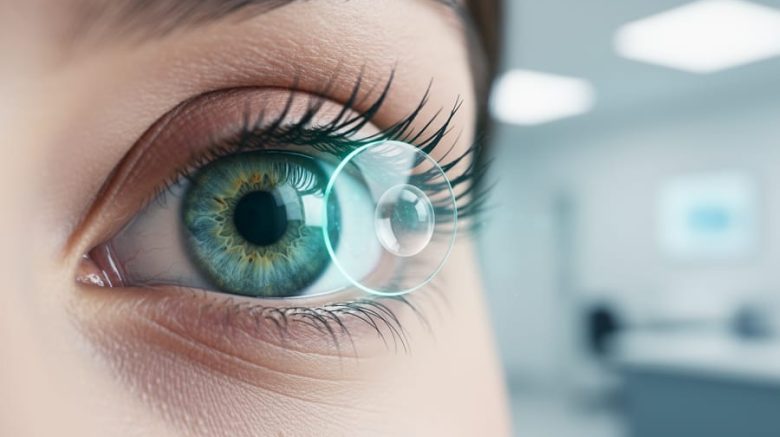
The eye is a complex sensory organ made of interconnected structures that work together to capture light and convert it into electrical signals your brain interprets as vision. Understanding how the eye’s anatomy and physiology function isn’t just academic curiosity. It explains why routine eye exams matter, how conditions like cataracts or glaucoma develop, and what happens when you focus on a sunset or read text on a screen.
Your eyes perform an extraordinary task every waking moment. Light enters through the transparent cornea, passes through the pupil (which adjusts size based on brightness), gets focused by the lens, and lands precisely on the retina at the back of your eye. There, millions of photoreceptor cells translate light into neural messages that travel via the optic nerve to your brain’s visual cortex. This entire process happens in milliseconds, allowing you to navigate your world with remarkable precision.
Each component of the eye serves a specific role. The cornea provides most of your eye’s focusing power. The iris controls light entry. The lens fine-tunes focus for near and far objects. The retina’s rods detect light and darkness while cones process color. Even the vitreous gel inside your eye maintains its shape and optical clarity.
This article breaks down the eye’s anatomy structure by structure, explains the step-by-step physiology of vision, and shows you how this knowledge applies to protecting your sight and recognizing when something needs professional attention.
What Eye Anatomy and Physiology Means
Eye anatomy and physiology are two complementary ways of understanding how your eyes work. Anatomy refers to the physical structures themselves, the cornea, lens, retina, and all the other parts you can identify and describe. Physiology, on the other hand, explains what those structures actually do and how they work together to create the vision you rely on every day.
- Anatomy
- The study of the eye’s physical structures and their arrangement, including tissues, layers, and components like the cornea, iris, and retina.
- Physiology
- The study of how eye structures function and interact to produce vision, from light entry to signal transmission to the brain.
- Vision
- The process by which your eyes and brain work together to interpret light and create the images you see.
- Refraction
- The bending of light rays as they pass through the cornea and lens to focus precisely on the retina.
- Photoreception
- The conversion of light into electrical signals by specialized cells in the retina, which are then sent to the brain.
Understanding both anatomy and physiology matters because knowing what your eye looks like is only half the picture. To truly grasp how conditions develop, why treatments work, or what symptoms mean, you need to understand the interplay between structure and function. A clouded lens (anatomy) causes blurred vision because it disrupts light focusing (physiology). When you comprehend both elements, you’re better equipped to recognize problems early and make informed decisions about your eye health.
The Main Components of Your Eye
The Outer Layer: Protection and Light Entry
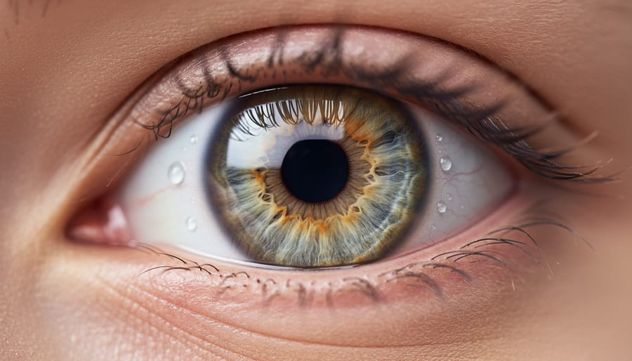
The eye’s outer layer forms a tough, continuous shell that shields delicate internal structures while allowing light to enter. This protective coating consists of two distinct regions working together.
The cornea covers the front of your eye, the clear, dome-shaped window you can see when you look in a mirror. Despite being transparent, it’s remarkably strong, made of five layers of specialized cells and proteins arranged in a precise pattern that maintains clarity while providing protection. The cornea does more than guard your eye; it bends incoming light rays to begin the focusing process, handling about two-thirds of your eye’s total focusing power.
The sclera forms the white part surrounding the cornea, extending around the entire eyeball. This opaque, fibrous tissue is tougher than the cornea and maintains your eye’s spherical shape against internal pressure. The sclera’s dense collagen fibers create a protective barrier against injury while providing attachment points for the muscles that move your eye.
Together, these structures create a sealed compartment that keeps your eye properly pressurized and shaped, essential for clear vision and protecting the sensitive tissues inside.
The Middle Layer: Blood Supply and Focus
The middle layer of your eye, called the uvea, consists of three connected structures that regulate light and keep your eye nourished. Think of it as the eye’s control and supply system, it manages how much light enters, adjusts focus, and delivers blood to fuel all that visual work.
The most visible part is the iris, the colored ring around your pupil. It’s actually a muscular diaphragm with an opening in the center that expands and contracts like a camera aperture. In bright conditions, muscles in the iris squeeze the pupil down to a pinpoint; in dim light, they relax and let the pupil widen to capture more available light. This automatic adjustment happens constantly without you thinking about it.
Behind the iris sits the ciliary body, a ring of muscle tissue with a less glamorous but crucial job. It produces the aqueous humor that fills the front of your eye and maintains proper pressure. More importantly, it controls your lens through tiny suspensory ligaments. When you look at something close, ciliary muscles contract and let the lens bulge for near focus. When you gaze into the distance, these muscles relax and flatten the lens out.
The choroid forms the back portion of this middle layer, a dense network of blood vessels sandwiched between the sclera and retina. It supplies oxygen and nutrients to the outer retinal layers and absorbs stray light that could otherwise scatter inside the eye and blur your vision.
The Inner Layer: Where Vision Begins
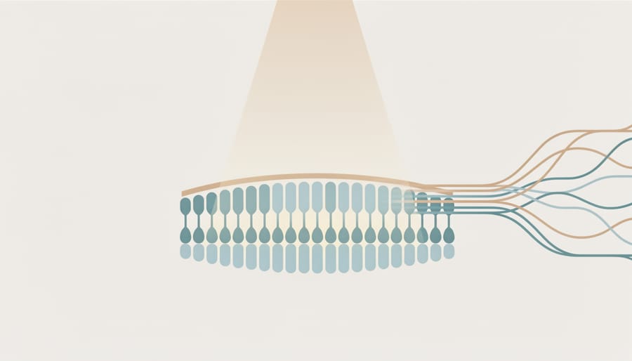
The retina is the innermost layer of your eye where the magic of vision actually starts. Think of it as the eye’s light-sensitive screen, a thin sheet of neural tissue lining the back of your eyeball that captures incoming light and transforms it into signals your brain can understand.
At the heart of the retina are photoreceptor cells, specialized neurons that perform the crucial job of converting light into electrical impulses. You have two types: rods and cones. Rods, numbering around 120 million, work best in dim conditions and handle your peripheral vision and ability to see at night. Cones, about 6 million in total, concentrate in the central retina and enable you to see color and fine detail in brighter light.
The macula is a small, oval yellowish area near the center of your retina, measuring about 5 millimeters across. It’s responsible for your sharpest, most detailed vision, the kind you use when reading, recognizing faces, or threading a needle.
Right at the macula’s center sits the fovea, a tiny pit less than a millimeter wide that contains the highest concentration of cone cells and zero rods. When you look directly at something, you’re positioning that image onto your fovea for maximum clarity. This pinpoint area gives you your clearest, most color-accurate vision, which is why damage to the fovea or macula dramatically affects your ability to see fine details.
Supporting Structures
Beyond the three tissue layers, your eye depends on several critical supporting structures that work behind the scenes to create clear vision. These components don’t form layers themselves, instead they fill spaces, focus incoming light, and connect your eye to your brain’s visual processing centers.
The lens sits just behind your iris, suspended by tiny fibers called zonules. This transparent, flexible structure fine-tunes focus by changing shape, flattening for distant objects and thickening for close-up work. Unlike the fixed cornea, your lens adjusts constantly as you shift your gaze between your phone screen and the horizon.
Two clear fluids maintain your eye’s shape and internal pressure. Aqueous humor fills the front chamber between your cornea and lens, continuously produced and drained to keep pressure stable. Vitreous humora gel-like substance, occupies the large cavity behind the lens, giving your eyeball its round form and keeping the retina pressed against the back wall.
The optic nerve serves as your eye’s information highway. This bundle of over one million nerve fibers exits through the back of your eye, carrying electrical signals from retinal cells to your brain’s visual cortex. Damage here disrupts the entire communication pathway, which is why conditions affecting the optic nerve can cause irreversible vision loss.
- Lens: adjustable focusing element that changes shape for near and far vision
- Aqueous humor: circulating fluid that nourishes front structures and regulates pressure
- Vitreous humor: gel filling that maintains eye shape and supports retinal position
- Optic nerve: nerve fiber bundle transmitting visual signals to the brain
Each supporting structure has a specialized maintenance requirement. Your lens depends on protein clarity, when those proteins clump, cataracts form. The aqueous humor’s drainage system must stay open to prevent glaucoma’s pressure buildup. The vitreous gel naturally liquefies with age, sometimes causing harmless floaters. And your optic nerve needs steady blood flow to keep transmitting signals reliably, anything that compromises circulation or compresses these fibers threatens your sight.
How Your Eye Creates Vision: The Process Step by Step
Light Refraction and Focus
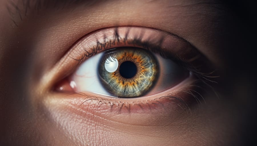
Vision starts the moment light enters your eye, but that light needs precise redirection to create a clear image. Your cornea handles about two-thirds of this light-bending work. Its curved, transparent surface refracts incoming light rays immediately, starting the focusing process before light travels deeper into the eye. The lens, sitting just behind the iris, contributes the remaining one-third of focusing power and provides something your cornea cannot: adjustability.
When you shift your gaze from a distant mountain to your phone screen, your lens changes shape through a process called accommodation. Tiny ciliary muscles surrounding the lens contract to make it thicker and more curved for close objects, increasing its refractive power. For distant objects, these muscles relax, allowing the lens to flatten and reduce its focusing strength. This dynamic adjustment happens automatically and constantly as you navigate your visual environment.
The goal of all this refraction is simple: converge light rays at exactly the right spot on your retina. When the cornea and lens work in harmony, light focuses precisely on the retina’s photoreceptor layer, producing sharp vision. If light focuses in front of the retina or behind it, as happens with nearsightedness or farsightedness, images appear blurred, which is where vision correction through glasses, contacts, or surgery becomes necessary to restore proper focus.
From Light to Electrical Signals
Once focused light hits your retina, something remarkable happens: specialized cells called photoreceptors transform light into the language your brain understands, electrical signals. This conversion is photoreception, the foundation of everything you see.
Your retina contains two types of photoreceptors, each designed for different vision tasks. Rods number around 120 million per eye and excel at detecting dim light and movement in your peripheral vision. They’re why you can navigate a dark room and spot motion from the corner of your eye, though they don’t detect color. Cones, numbering about 6 million, concentrate in your central retina (especially the fovea) and specialize in sharp detail and color vision. Three cone types respond to different wavelengths, red, green, and blue, giving you the full color spectrum you experience.
When light photons strike these cells, they trigger a chemical cascade. A light-sensitive molecule called rhodopsin (in rods) or photopsin (in cones) changes shape, initiating a series of reactions that generate an electrical impulse. This signal travels through several retinal cell layers, first to bipolar cells, then to ganglion cells whose long fibers bundle together to form the optic nerve. The optic nerve carries ganglion signals away from your eye toward your brain’s visual processing centers.
This entire conversion happens in milliseconds, with your retina constantly adjusting its sensitivity based on lighting conditions, a sophisticated biological process that makes every visual moment possible.
Transmitting Visual Information
Once nerve impulses leave the retina, they travel along roughly 1.2 million nerve fibers bundled into the optic nerve, a cable about the thickness of a pencil that exits the back of each eye. These signals first converge at the optic chiasm, where fibers from the nasal half of each retina cross to the opposite side of the brain. This crossing ensures that each side of your brain processes information from both eyes, creating a unified visual field.
From there, most signals route through the lateral geniculate nucleus, a relay station in the thalamus that filters and organizes visual information by type. Finally, impulses reach the visual cortex at the back of your brain, where specialized neurons decode color, shape, motion, and depth. Different cortical areas handle different tasks: one region recognizes faces, another interprets written words, and others coordinate eye movements.
Your brain doesn’t just passively receive these signals. It actively compares them against stored memories, fills in blind spots, and combines visual data with input from other senses, which is why you can recognize a friend’s face instantly or know that a sizzling sound matches the steak you see cooking. This integration happens in milliseconds, creating the seamless experience you call sight.
How Eye Anatomy Supports Different Vision Functions
Color and Detail Vision
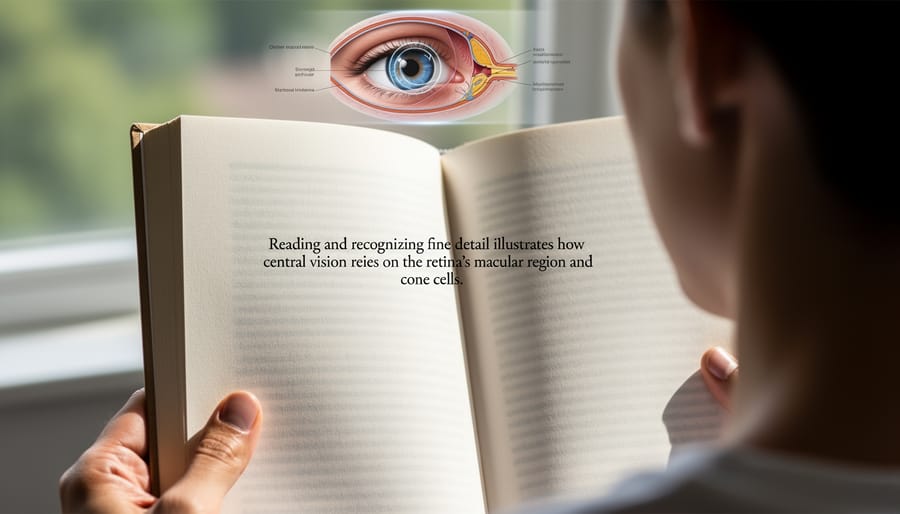
Your ability to read these words, distinguish the blue of a hyperlink from black text, and focus on a single letter depends on a tiny spot in your retina called the macula. This 5-millimeter region near the center of your retina packs the highest concentration of cone cells, specialized photoreceptors responsible for color vision and fine detail.
The macula contains three types of cones, each sensitive to different wavelengths: red, green, or blue. When light hits these cells, they work together to create the full spectrum of colors you perceive. At the macula’s center sits the fovea, an even smaller depression where cone density reaches its peak and supporting layers thin out to allow maximum light transmission. This tiny area provides your sharpest vision, what you use for reading, recognizing faces, threading a needle, or examining anything requiring precision.
When you look directly at an object, your eyes automatically rotate so its image falls on the fovea, giving you crystal-clear central vision while peripheral details remain fuzzy. This anatomical design explains why damage to the macula, as occurs in macular degeneration, creates a blind spot right where you need vision most.
Low-Light and Peripheral Vision
Your ability to navigate a dimly lit room or catch movement in the corner of your eye depends entirely on specialized structures in your peripheral retina. While cone cells dominate the central macula, rod cells, approximately 120 million of them, populate the outer regions of your retina and operate quite differently.
Rods excel in low-light conditions because they’re extraordinarily sensitive to single photons. They don’t detect color, which is why everything looks grayscale at night, but they respond to minimal light levels that would leave cones inactive. This explains why you see better in the dark using your peripheral vision rather than looking directly at something.
The peripheral retina also acts as your motion detection system. Because rods respond quickly to changes in light patterns, they alert your brain to movement before you consciously turn to look. This spatial awareness was crucial for survival throughout human evolution, warning us of approaching threats.
When you move from bright sunlight into a dark theater, your rods need about 20 to 30 minutes to fully adapt, gradually becoming more sensitive as a light-absorbing pigment called rhodopsin regenerates in these cells.
Eye Movement and Coordination
Six small extraocular muscles attached to each eye’s outer surface allow you to track moving objects, shift your gaze, and maintain alignment between both eyes. These muscles work in coordinated pairs, when one contracts, its opposing partner relaxes, enabling smooth, precise movements in all directions. Your eyes perform rapid movements called saccades when reading or scanning a scene, slower tracking movements when following a moving car, and vergence movements when shifting focus between near and far objects.
This muscular coordination extends beyond the eyes themselves. The brain integrates visual input with motor control, creating the seamless hand-eye coordination you use when catching a ball or threading a needle. Binocular vision, the ability to fuse images from both eyes into a single three-dimensional perception, depends entirely on precise muscle control. When these muscles don’t work together properly, you might experience double vision, eye strain, or difficulty judging distances. Regular use strengthens this coordination, which is why activities requiring visual tracking can improve overall eye-muscle function.
Why Understanding Eye Anatomy Matters for Your Health
Knowing how your eye is built transforms you from a passive patient into an informed participant in your own care. When your ophthalmologist mentions that pressure is building in your anterior chamber, you’ll understand they’re talking about the space between your cornea and iris, and why that matters for glaucoma risk. When you notice halos around lights at night, you’ll recognize this might signal corneal swelling or early cataract formation in your lens.
This anatomical literacy helps you catch problems early. A sudden increase in floaters paired with flashes of light becomes more alarming when you understand these could indicate retinal detachment, a tear in the tissue that converts light to vision. Blurry central vision takes on new urgency when you know it might affect your macula, the retinal region responsible for reading and recognizing faces.
Understanding structure also clarifies why certain treatments work. Glaucoma medications reduce aqueous humor production or improve its drainage because you now know this fluid maintains your eye’s shape and pressure. Cataract surgery makes sense as a lens replacement procedure. Anti-VEGF injections for macular degeneration target abnormal blood vessel growth in the choroid layer beneath your retina.
Your daily choices gain new meaning too. Wearing UV-protective sunglasses isn’t just general advice, it shields your lens and retina from cumulative damage. Maintaining healthy blood pressure protects the delicate vessels in your choroid and retina. Managing blood sugar levels preserves the tiny capillaries feeding your macula. Learning about eye nutrition shows how specific nutrients support photoreceptor health and retinal function.
Knowledge of common eye conditions and their anatomical basis makes you a better advocate for your vision. Combined with preventive eye care this understanding helps you spot warning signs and take action before minor issues become major problems.
Common Questions About Eye Anatomy and Physiology
Why do my pupils get bigger and smaller? Your pupils adjust automatically to control how much light enters your eye, widening in dim conditions to gather more light and shrinking in bright environments to prevent overexposure. The iris muscles handle this adjustment through the pupillary light reflex, which happens in less than half a second. Certain medications, emotions, and neurological signals can also affect pupil size.
What causes those floating spots I sometimes see? Floaters appear when tiny clumps of gel or cells inside the vitreous humor cast shadows on your retina. As you age, the vitreous becomes more liquid and can form these harmless strands, which drift around when you move your eyes. While most floaters are normal, a sudden increase in them or flashes of light should prompt an immediate eye exam, as these can signal retinal problems.
How do my eyes actually focus on objects at different distances? Your eyes focus through accommodation, the ciliary muscles contract to make your lens thicker for near vision and relax to flatten it for distant vision. This happens automatically when you shift your gaze from your phone to the road ahead. After age 40, the lens becomes less flexible, which explains why many people start needing reading glasses for close-up tasks.
Can I see without my cornea?
No, your cornea provides roughly 65% to 75% of your eye’s focusing power. Without it, light rays wouldn’t bend correctly to reach your retina, leaving you with severely blurred vision or functional blindness.
Why do my eyes get red sometimes?
Redness occurs when blood vessels in your sclera and conjunctiva expand due to irritation, dryness, fatigue, or infection. The visible redness is actually increased blood flow bringing immune cells and oxygen to address whatever is bothering your eye.
Does eye anatomy change as I age?
Yes, several structures change naturally with age. Your lens hardens and loses flexibility (presbyopia), your vitreous liquefies, your pupils get smaller, and your tear production may decrease. These changes are normal but increase the importance of regular eye exams.
Why are some people’s eyes different colors?
Eye color comes from the amount and type of melanin pigment in your iris. Brown eyes have more melanin, blue eyes have less, and green or hazel eyes fall in between, the structure of the iris itself is the same regardless of color.
These anatomical quirks and changes are part of how your eyes adapt to your environment and age. Understanding them helps you recognize what’s normal variation versus what deserves professional attention.
Understanding how your eye is built and how it works gives you real power over your eye health. When you know that the macula handles your central vision or that the optic nerve transmits signals to your brain, you can better recognize warning signs like blurry spots or sudden vision changes. This knowledge helps you ask the right questions during appointments and make informed decisions about treatments or lifestyle changes.
Regular eye exams become more meaningful when you understand what your eye doctor is checking. They’re not just testing how well you see, they’re examining the health of your cornea, monitoring pressure that could affect your optic nerve, and looking at your retina for early signs of damage. These structures work hard every second of every day, and routine monitoring helps catch problems before they affect your vision.
Your eyes are remarkably complex organs with an elegant design honed over millions of years of evolution. Despite their delicate structures, they’re also resilient, constantly adapting to changing light conditions and repairing minor damage. Respecting that complexity by protecting your eyes, nourishing them properly, and getting professional care when needed helps ensure they serve you well for decades to come.