
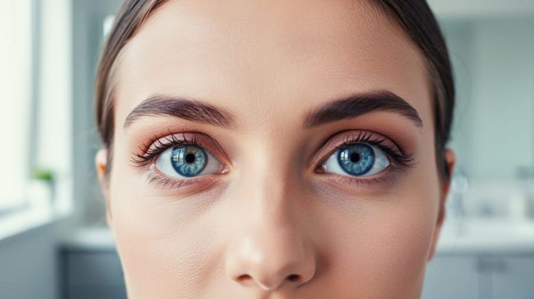
Eye conditions reveal themselves through distinctive visual changes that range from subtle color shifts to dramatic alterations in the appearance of the eyes themselves. Understanding what these conditions look like helps you recognize warning signs early and know when professional care is needed.
Your eyes offer visible clues about their health. Many common conditions produce telltale signs you can spot in a mirror or notice in someone else: the yellowing of jaundice, the cloudiness of cataracts, the redness of conjunctivitis, or the uneven pupils that signal nerve damage. Some changes develop gradually over years, while others appear suddenly within hours.
The challenge is knowing which visual symptoms matter. Not every red eye requires emergency attention, but certain patterns demand immediate care. A bright red spot might be a harmless broken blood vessel that resolves on its own, while a hazy cornea could indicate serious infection requiring same-day treatment.
This guide walks you through what different eye conditions actually look like, organized by the type of visible change they produce. You’ll learn to identify the physical characteristics of common conditions, understand what’s happening beneath the surface to cause these appearances, and recognize which visual symptoms warrant a call to your eye care provider versus which can wait for your next routine exam.
What Eye Conditions Are and What They Look Like
Eye conditions are abnormalities that affect the eyes’ structure, function, or appearance. These can range from minor irritations that resolve on their own to serious diseases requiring immediate medical attention. Some conditions develop gradually over years, while others appear suddenly within hours or days.
Understanding what eye conditions look like starts with recognizing two categories of visible changes. External signs are alterations you or others can observe when looking at your eyes, redness, swelling, discharge, unusual coloring, or structural changes. Internal symptoms affect what you see or feel but may not be visible to others, such as blurred vision, floaters, or light sensitivity. Many conditions produce both types of indicators.
- Eye Condition
- Any abnormality affecting the eye’s structure, function, or appearance, from temporary irritations to chronic diseases.
- External Signs
- Visible changes others can see when looking at your eyes, including color alterations, swelling, discharge, or structural abnormalities.
- Internal Symptoms
- Changes in vision or eye sensation that you experience but others cannot observe directly, such as blurriness or pain.
- Visual Indicators
- Observable clues that signal a potential eye problem, combining what you see in the mirror with what you experience visually.
The good news is that many eye conditions have recognizable visual patterns. A bright red spot suggests subconjunctival hemorrhage, while a milky pupil often indicates cataracts. Crusty eyelid edges point to blepharitis, and a drooping lid signals ptosis. Learning these patterns helps you identify when something needs attention and communicate symptoms clearly to your eye care provider. Not every condition is immediately visible, but most produce signs you can detect with careful observation.
How Eye Conditions Affect What You See and How Your Eyes Look
Eye conditions alter both what you experience internally and what others notice about your appearance through five primary biological mechanisms. Understanding these processes helps you recognize potential problems early and communicate symptoms effectively to eye care professionals.
Inflammation causes visible redness, swelling, and discomfort when blood vessels in the eye or eyelid dilate in response to irritation, injury, or infection. The white part of your eye may appear pink or bloodshot, while eyelids can become puffy and tender. Inflammation often produces a gritty or burning sensation that affects how clearly you see.
Fluid buildup creates swelling around the eyes and within eye structures. When fluid accumulates in the eyelids, they appear bloated and heavy. Inside the eye, fluid retention can cloud the cornea or lens, making your vision hazy or creating halos around lights. This mechanism explains why some conditions cause both visible puffiness and simultaneous vision changes.
Structural damage or changes physically alter eye anatomy in ways you and others can see. A cataract clouds the normally clear lens, creating a milky or yellowish appearance in the pupil area. Corneal scarring might look like white or gray patches on the eye’s surface. These structural shifts typically correspond directly to what you experience visually, clouded structures mean clouded vision.
Infection produces discharge, crusting, and color changes as your body fights bacteria, viruses, or other pathogens. Yellow or green discharge that crusts overnight signals bacterial infection, while watery, clear discharge often indicates viral causes. The affected eye typically appears red and may stick shut upon waking.
Vascular and circulation issues make blood vessels more visible or cause bleeding within the eye. Dilated vessels create the classic bloodshot appearance, while a broken blood vessel produces a bright red patch on the white of the eye. Poor circulation can contribute to color changes, including yellowing or paleness of tissues around the eyes.
These mechanisms rarely work in isolation. Many conditions trigger multiple processes simultaneously, conjunctivitis combines infection, inflammation, and discharge, while glaucoma might involve both fluid buildup and structural changes to the optic nerve that aren’t visible externally but profoundly affect peripheral vision.
Common Eye Conditions and Their Visual Appearance
Red Eye Conditions
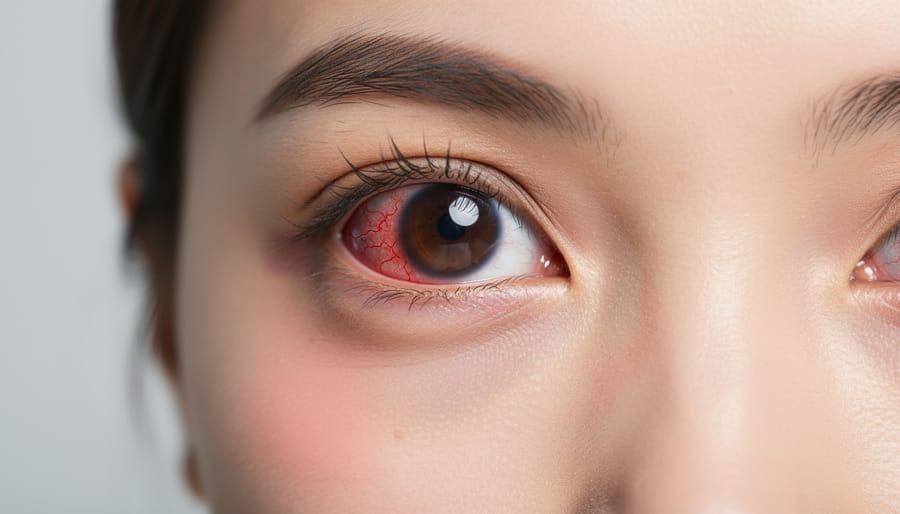
Red eyes are one of the most common visible symptoms that prompt concern, but not all redness means the same thing. The pattern, intensity, and accompanying features help distinguish between different conditions.
Conjunctivitis (Pink Eye)
Conjunctivitis creates a distinctive pink or red color that spreads across the white of the eye, often starting in one eye before affecting both. The redness typically appears diffuse rather than concentrated in one spot. You’ll notice discharge that can be watery, thick, or crusty, depending on whether the cause is viral, bacterial, or allergic. Bacterial conjunctivitis often produces yellow or greenish discharge that crusts overnight, making it hard to open your eyes in the morning. Viral cases tend to be more watery with less crusting. The eyelids may look swollen and feel gritty, as though something’s stuck under them.
Subconjunctival Hemorrhage
This condition looks dramatic but usually isn’t serious. A bright red patch appears on the white of the eye where a tiny blood vessel has broken, creating a blood-filled area with sharp, defined edges. Unlike the diffuse redness of conjunctivitis, this blood spot has clear boundaries and doesn’t spread across the entire eye. There’s no discharge, and the blood doesn’t move or smear. It may expand slightly over the first day but then gradually fades to yellow as it heals over one to three weeks.
Dry Eye Syndrome
Dry eye creates red and scratchy eyes with a threadlike pattern of redness that’s more prominent near the corners and edges. The whites may look slightly bloodshot with visible small vessels rather than an overall pink wash. You might see stringy mucus in the corners, and the eye surface can appear dull rather than glossy.
- Conjunctivitis: Diffuse pink/red color with discharge and crusty lashes
- Subconjunctival hemorrhage: Sharply defined bright red blood patch with no discharge
- Dry eye: Threadlike vessel pattern with dull surface and stringy mucus
These visual differences matter because each condition requires different treatment approaches. While conjunctivitis and dry eye are common elderly eye conditions they can affect anyone at any age. Recognizing which type of redness you’re seeing helps you decide whether home care is appropriate or professional attention is needed.
Structural and Shape Changes
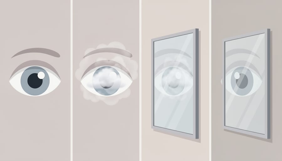
Structural changes to the eye often catch your attention when you look in the mirror because they alter the familiar shape or appearance of your eyes.
Cataracts create a cloudy, milky appearance in the pupil that you might first notice in photographs where one eye reflects the flash differently than the other. As they progress, the normally black pupil takes on a grayish, yellowish, or whitish haze, similar to looking through a foggy window. You’ll experience vision changes long before others notice the cloudiness, colors may appear faded, lights develop halos, and reading becomes difficult despite good lighting. Advanced cataracts become visible to others as a distinct white or amber cloudiness behind the pupil.
Ptosis, or eyelid drooping, changes the symmetry of your face. One or both upper eyelids may hang lower than normal, partially covering the pupil and creating an asymmetrical appearance. In the mirror, you’ll see less of your iris on the affected side. Severe ptosis can block your upper field of vision, making you tilt your head back to see properly or raise your eyebrows constantly to compensate.
Styes appear as red, painful bumps along your eyelid edge, resembling a pimple. They typically point outward and may develop a small yellow spot of pus at the tip. Chalazia are larger, firmer lumps that usually form away from the lid edge. They feel like a hard pea under the skin and may cause the entire eyelid to swell. While styes are tender, chalazia often don’t hurt unless they grow large enough to press on the eyeball.
Corneal conditions can create visible surface changes. Corneal abrasions might not look obvious externally but cause intense pain, tearing, and light sensitivity. Keratoconus gradually changes the cornea’s shape from round to cone-like, which you might notice as increasing distortion in your vision rather than an obvious external change.
Color and Clarity Changes
The whites of your eyes, called the sclera, should appear bright white or very slightly off-white with fine blood vessels visible on close inspection. Significant color changes often signal underlying conditions worth investigating.
Yellow Discoloration
When the sclera turns yellow or develops a yellowish tint, it typically indicates jaundice caused by elevated bilirubin levels in the bloodstream. This yellowing usually appears evenly across the white portions of both eyes and often accompanies yellowing of the skin. Liver conditions, bile duct problems, or blood disorders commonly cause this change. The yellowing becomes more noticeable in natural light and contrasts sharply with the darker iris.
Gray or White Rings
Arcus senilis appears as a gray, white, or bluish ring around the outer edge of the cornea. This ring develops gradually as lipid deposits accumulate with age and becomes increasingly common after 60. The ring itself doesn’t affect vision and represents a normal aging change in most older adults. In younger individuals under 40, however, a similar ring (arcus juvenilis) may indicate high cholesterol or lipid disorders requiring medical evaluation.
Cloudy or Hazy Appearance
When eyes develop a cloudy, filmy, or hazy look, several conditions might be responsible. Cataracts create cloudiness within the pupil area that appears grayish-white or milky, often compared to looking through a foggy window. Corneal edema from injury or disease produces generalized haziness across the entire cornea. Severe dry eye can create a dull surface appearance rather than the normal glossy sheen. These changes typically develop gradually rather than appearing suddenly.
Swelling and Inflammation
Swelling and inflammation around the eyes create distinct visual patterns that help identify specific conditions. The location, severity, and accompanying symptoms of puffiness provide important clues about what’s causing the problem.
Blepharitis produces inflammation along the eyelid margins where the lashes emerge. You’ll notice red, swollen eyelid edges with visible crusting or flaking, especially after waking up. The lashes may have small white or yellowish scales clinging to their bases, resembling dandruff. Eyes often appear greasy or irritated, and the lid margins may look thickened. This chronic condition typically affects both eyes, though one side can be worse than the other.
Allergic reactions cause rapid, sometimes dramatic swelling of the eyelids and surrounding tissues. The lids puff up and may look shiny or tight, sometimes swelling so much that the eye appears nearly closed. The skin takes on a pale or slightly bluish tint rather than the angry red of infection. You might see fluid retention creating a watery appearance beneath the lower lids. Allergic swelling often affects both eyes symmetrically and can extend to the cheeks and under-eye conditions.
Uveitis, while primarily affecting internal eye structures, sometimes produces visible external signs. The eye appears red, particularly around the iris, with a deep, dusky redness rather than the bright pink of conjunctivitis. You may notice the pupil appears smaller or irregularly shaped compared to the other eye. The eyelids can show mild swelling, and the eye often looks watery with light sensitivity causing squinting.
Different swelling patterns signal different causes: localized bumps suggest styes or chalazia, generalized puffiness indicates allergic or systemic issues, and eyelid margin inflammation points to blepharitis or infection.
When and How to Use This Visual Information
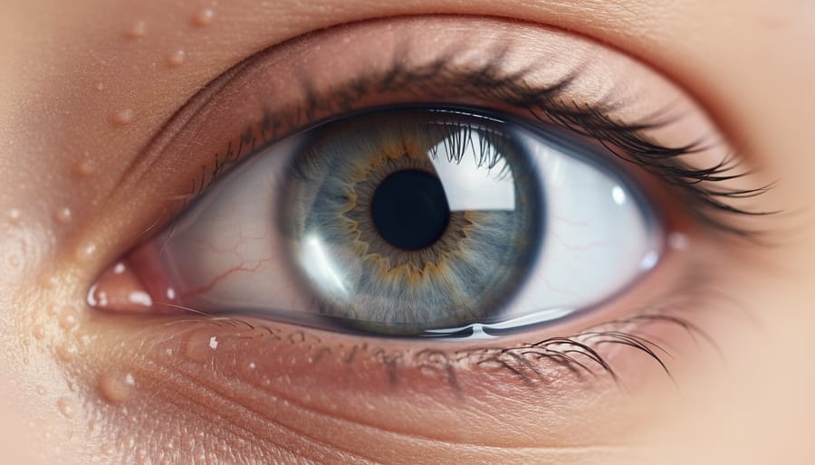
Knowing what eye conditions look like gives you a practical tool for monitoring your health, but using this information effectively requires a balanced approach. Check your eyes regularly in good natural light using a clean mirror. Look for changes in color, new redness, unusual discharge, swelling, or bumps. Notice whether both eyes look the same or if one differs from the other.
Compare what you see today against your normal baseline. Most people have slight variations between eyes, tiny visible blood vessels, or minor asymmetries that are perfectly normal. What matters is change: a new bright red spot, sudden cloudiness, progressive yellowing, or swelling that wasn’t there before.
Document changes before your appointment. Take photos in consistent lighting from the same distance, capturing both eyes for comparison. Note when symptoms started, whether they’re getting worse, and what makes them better or hurt more. This timeline helps your eye care provider understand patterns and urgency.
Some visual changes demand immediate attention. Seek same-day or emergency care for sudden vision loss, severe eye pain, pupils that differ in size, curtain-like shadows across your vision, or bright red blood filling the white of your eye after trauma. Flashes of light with new floaters, sudden double vision, or bulging eyes also warrant urgent evaluation.
Other symptoms need prompt but not emergency care within a few days. Schedule an appointment soon for persistent redness lasting more than two days, discharge that returns after cleaning, gradual vision changes, new floaters without flashes, or eyelid swelling that limits your eye opening.
Routine monitoring fits into your broader preventive eye care strategy. Annual exams catch conditions before visible symptoms appear, while between visits you watch for changes. Early recognition improves outcomes for many conditions. Catching cataracts early allows time to plan vision correction when you’re ready. Noticing dry eye symptoms early means starting treatment before corneal damage occurs.
Remember that workplace eye safety matters too. If you work with tools, chemicals, or flying debris, wear safety glasses regardless of what you currently see. Prevention beats treatment every time.
Use visual knowledge as one piece of information, not a replacement for professional diagnosis. Your eye care provider has specialized equipment to see structures you can’t examine yourself and training to distinguish serious conditions from harmless variations.
Expert Guidance on Recognizing Eye Conditions
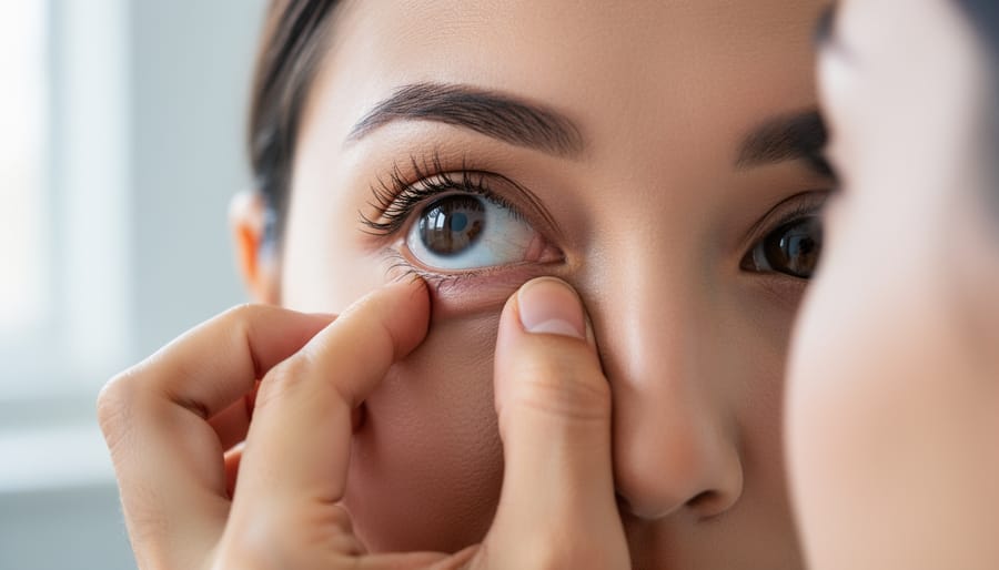
Recognizing changes in your eyes starts with proper examination technique. Stand in front of a well-lit mirror in natural daylight or under bright white light, which reveals color changes and subtle symptoms that dim lighting might hide. Gently pull down your lower lid to check the inner surface for redness or discharge, then look upward while examining the white part of your eye for discoloration or blood vessels. Check both eyes separately, comparing them for asymmetry.
If you notice changes, photograph them for your eye care provider. Take close-up images in good lighting from multiple angles, capturing the overall eye and any specific areas of concern. Include photos with your eyelids open and gently pulled down to show the conjunctiva. These images help specialists track progression between appointments and provide context for your symptoms.
Understand that self-examination has limits. You can’t see your retina, optic nerve, or internal eye pressure. Many serious conditions like glaucoma and macular degeneration develop without visible external signs. What looks like simple redness might mask deeper inflammation only specialized equipment can detect.
Eye care professionals use tools beyond visual inspection: slit lamps magnify eye structures under high-intensity light, tonometers measure internal pressure, and ophthalmoscopes examine the retina and blood vessels inside your eye. They assess tear production, check peripheral vision, and evaluate how your pupils respond to light.
Seek professional examination if you experience sudden vision changes, persistent redness lasting more than two days, pain, light sensitivity, or new floaters and flashes. Regular comprehensive eye exams catch conditions before they produce visible symptoms. Combine self-monitoring with proper eye nutrition and routine professional care for complete eye health protection.
How What Do Eye Conditions Look Like Works
Recognizing eye conditions by appearance relies on understanding how the eye’s transparent and reflective structures reveal internal changes. The front of your eye acts like a window, when something goes wrong inside, it often shows through as color shifts, cloudiness, or swelling you can see from the outside.
The white part of your eye (sclera) contains blood vessels that dilate when irritated, creating visible redness. Clear structures like the cornea and lens become cloudy when proteins break down or debris accumulates. The conjunctiva, a thin membrane covering the white part, turns pink or red when inflamed and can develop yellow or white discharge that’s visible at the corners.
Fluid dynamics play a major role. When blood vessels break, you see bright red patches. Blocked oil glands create visible bumps along the eyelid margin. Excess fluid from inflammation causes puffiness you can observe in the mirror.
Your pupils also signal problems. They should appear black and round, but certain conditions make them look cloudy, irregular, or unequal in size. The iris color changes subtly with some diseases, shifting from clear blue to grayish or developing rust-colored spots.
These visible signs emerge because the eye’s normally transparent tissues lose clarity or because blood flow and drainage patterns change in response to disease.
How What Do Eye Conditions Look Like Is Used
Understanding what eye conditions look like serves several practical purposes in everyday healthcare. Patients use this knowledge to monitor their own eye health between routine exams, catching changes early when treatment is most effective. Parents rely on visual recognition to identify potential problems in children who may not articulate symptoms clearly, spotting a cloudy pupil or persistent eye redness prompts timely pediatric visits.
Caregivers working with elderly patients or those with cognitive challenges use appearance cues to advocate for needed care, recognizing when yellowing, swelling, or discharge indicates infection or other treatable conditions. Emergency department staff apply visual assessment to triage patients, distinguishing urgent conditions like acute angle-closure glaucoma from less critical issues.
Primary care physicians use appearance-based screening to determine when specialist referral is necessary. They might identify a suspicious growth, unexplained pupil changes, or inflammatory patterns requiring ophthalmologic evaluation.
Even eye care specialists benefit from patients who arrive with detailed observations and photos documenting symptom progression, this visual timeline helps pinpoint diagnosis and assess treatment response. The ability to recognize and describe what you’re seeing transforms vague concerns into actionable medical information.
Common Questions About Eye Condition Appearance
Knowing what to look for is helpful, but many people have similar questions about interpreting what they see. Understanding the limits of self-assessment and recognizing when professional evaluation is necessary can help you respond appropriately to eye changes.
Can I diagnose my own eye condition just by looking?
No, you can’t reliably diagnose eye conditions through self-examination alone. While you can recognize signs that warrant professional attention, many conditions with similar appearances have different causes requiring specific treatments, and some serious issues show minimal external changes.
What does a healthy eye look like?
A healthy eye typically has clear, white sclera with minimal visible blood vessels, a smooth cornea, symmetrical pupils that react to light, and clean eyelid margins without crusting or swelling. Some minor variations like small visible capillaries are normal for many people.
Are red eyes always a sign of something serious?
Red eyes aren’t always serious, they can result from minor irritation, allergies, dry air, or fatigue. However, redness accompanied by pain, vision changes, light sensitivity, or discharge warrants prompt evaluation to rule out infection or inflammation.
When should I photograph my eye symptoms for my doctor?
Photograph symptoms when they fluctuate throughout the day, appear intermittently, or are most prominent at specific times. Take clear, well-lit photos showing both eyes for comparison, and include images at different stages if the condition is changing.
The speed at which eye symptoms develop matters significantly. Sudden changes, a bright red spot appearing within hours, rapid swelling, or abrupt vision loss, typically require urgent evaluation. Gradual changes over weeks or months, like slowly developing cloudiness or a growing bump, still need professional assessment but don’t usually constitute emergencies. Track how quickly symptoms progress, as this information helps your eye care provider determine the likely cause.
Age brings normal changes that can look concerning if you’re not expecting them. Arcus senilis, that grayish ring around the cornea, becomes common after 60 and doesn’t affect vision. The conjunctiva may develop small, harmless yellow spots or visible blood vessels with age. However, don’t dismiss changes as “just aging” without verification, new floaters, persistent redness, or vision shifts deserve evaluation regardless of your age.
Recognizing what different eye conditions look like gives you valuable awareness, but it’s just the starting point. While visible symptoms like redness, swelling, or cloudiness can alert you to potential problems, they represent only part of the diagnostic picture. Eye care professionals use specialized equipment to examine internal structures, measure pressure, and detect conditions that show no outward signs.
Think of visual changes as your eyes’ way of flagging attention. Some symptoms, sudden vision loss, severe pain, or trauma, demand immediate care. Others develop gradually and need monitoring. The key is responding appropriately rather than dismissing changes or delaying professional evaluation.
Make eye monitoring part of your routine wellness. Check your eyes in good lighting, note any changes, and schedule regular comprehensive exams even when nothing seems wrong. Many serious conditions, including glaucoma and diabetic retinopathy, progress silently without visible external symptoms.
Your observations matter. When you can describe what you’ve noticed, when it started, how it’s changed, what makes it better or worse, you help your eye care provider reach an accurate diagnosis faster. Combine self-awareness with professional expertise, and you’re taking the best possible approach to protecting your vision for life.